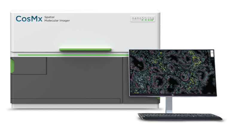
The CosMx SMI platform is the pioneering high-plex in-situ analysis tool that offers spatial multiomics for both FFPE and FF tissue samples at the cellular and subcellular levels. With up to 1,000 RNA and 64 validated protein analytes, CosMx SMI allows for quick quantification and visualization.
With the CosMx Spatial Molecular Imager (SMI), scientists can create comprehensive maps of individual cells within their natural environment. This technology enables researchers to gain a deeper understanding of various cell types, their behaviors and functions, ultimately enhancing our ability to interpret biological processes and diseases from a single experiment.
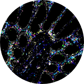
How does CosMx work?
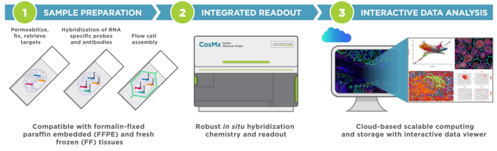
The CosMx SMI is a comprehensive system that features advanced cyclic fluorescent in-situ hybridization (FISH) chemistry, high-quality imaging output, interactive data analysis and visualization software. Its workflow is efficient and easy-to-use, and it seamlessly integrates with standard ISH protocols without requiring tissue expansion or clearing, cDNA synthesis, or amplification. With the CosMx SMI, you can achieve faster results from your samples.
Why choose CosMx?
The CosMx™ system is designed for spatial profiling of gene expression in tissues. It combines the capabilities of traditional histology with high-plex RNA profiling at single-cell resolution. Here are some advantages of the CosMx™ Spatial Molecular Imager:
Spatially Resolved Gene Expression: The CosMx™ system enables researchers to visualize gene expression patterns within intact tissue sections, providing spatial context for cellular behavior and interactions.
Single-Cell Resolution: It allows the identification and characterization of individual cells within a tissue, enabling a deeper understanding of cellular heterogeneity and complex biological processes.
High-Plex RNA Profiling: With the use of NanoString’s barcoding technology, the CosMx™ system can simultaneously measure the expression of hundreds of genes in a single tissue section, allowing comprehensive molecular profiling.
Multiplexed Imaging: The CosMx™ system enables the detection of multiple RNA targets within a single tissue section, providing a wealth of information on gene expression patterns and interactions.
Easy Workflow: The system offers an integrated workflow from tissue processing to data analysis, streamlining the experimental process and allowing researchers to focus on data interpretation and biological insights.
Heterogeneity, resolved
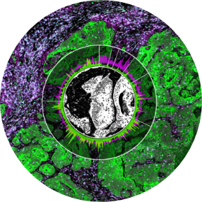
Spatially resolve tissues and cell populations with functional segmentation.
Detect more, without compromise.
Detect more of the transcriptome and proteome with the highest plex and sensitivity.
Consistent results, reliable answers.
Multi-sample analysis and cohort studies made easy with unmatched reproducibility and scalability.
Unlock your samples, with confidence.
Get proven, robust results from FFPE, FF tissues and TMAs using standard histology workflows.
Structure dictates function, think outside the box.
Profile functionally distinct cells and structures to get a complete picture of the biology that matters.
Analyse today, publish faster.
Don’t wait. Get publication- ready results faster with higher throughput and an integrated data analysis software.
CosMx Spatial Molecular Imager is the most flexible and robust spatial single-cell imaging platform for
Cell Atlas and Characterization
Define cell types, cell states, tissue microenvironment phenotypes, and gene expression networks within spatial context.
Biomarker Discovery
Reveal differential gene expression and pathways in the same cell types depending on their spatial location.
Disease State
Visualize and quantify molecular (RNA / protein) and cellular organizational changes in a tissue.
Tissue Microenvironment
Understand cellular neighborhoods by examining individual cells and their interacting neighboring cells.
Ligand-Receptor Interaction
Analyze expression and interactions of up to 100 classic ligand-receptor pairs between interacting cells.
Spatial Biomarkers
Quantify change in gene expression based on treatment and identify single-cell subcellular biomarkers with spatial context.
Panel Assays
Flexible options to cover your research needs
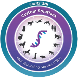
CosMx Custom Solutions Panel
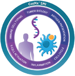
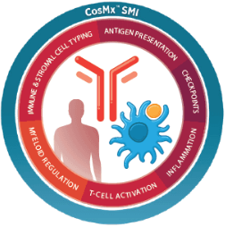
CosMx Human Universal Cell Characterization Panel (RNA, 1000 Plex)
CosMx Human Immuno-oncology Panel (RNA, 100 Plex)
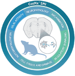
CosMx Mouse Neuroscience Panel (RNA, 1000 Plex)
CosMx Human Immuno-oncology Panel (Protein, 64 Plex)
Analysis
LifeStrands provides all customers a one-stop solution to all their spatial needs by having an in-house bioinformatician to aid you with their research.
The only cloud-based, fully-integrated informatics platform for spatial biology. Will be used for all Nanostring technologies. With advanced analytics enabling global data sharing and collaboration, AtoMx SIP helps researchers analyze large amounts of spatial multiomics data anytime, anywhere.
NanoString Spatial Platform Integration
Stream image and count files seamlessly from CoxMx SMI and GeoMx DSP into AtoMx SIP.
Data Management
Securely store, manage, and collaborate spatial multiomics data around the globe.
Analyze & Visualize Spatial Data
Perform secondary analysis and tertiary analysis with preconfigured modules and data analysis pipelines.
Customer Analysis
Edit pre-built modules and pipelines OR build custom module and pipelines to analyze spatial data to fit your needs.*
Security & Compliance
Single Sign-On authentication backed by standard-compliant data encryption for data-in0transit and data-at-rest through Amazon Web Services.
Our Technologies
Nanostring (GeoMx)
NanoString’s GeoMx Digital Spatial Profiler (DSP) is a technology that enables comprehensive spatial and molecular analysis of RNA and protein in tissue samples on glass slides.
Nanostring (CosMx)
CosMx SMI is the first high-plex in situ analysis platform to provide spatial multiomics with formalin-fixed paraffin-embedded (FFPE) and fresh frozen (FF) tissue samples at cellular and subcellular resolution.